
Xanh99 – Cổng game bài đổi thưởng có nhiều người chơi nhất 2025 – 2024
Xanh99 là một cổng game bài đổi thưởng uy tín với 7 năm thâm niên trong ngành. Cổng game này nổi bật với đồ họa chất lượng và giao diện thân thiện với người dùng, mang lại trải nghiệm tốt cho người chơi. Nhờ vào nhiều chương trình khuyến mãi hấp dẫn và tri ân khách hàng thường xuyên, Xanh99 đã xây dựng được sự tin tưởng từ cộng đồng game thủ.
Cổng game cũng sở hữu hệ thống giao dịch nhanh chóng với đa dạng phương thức thanh toán, giúp người chơi thực hiện nạp rút dễ dàng và thuận tiện.
Link tải Xanh99
xanh99.com/apk
xanh99.com/ios
xanh99.com/android
xanh99.com/webgame
Tên miền chính thức Xanh99
xanh99.com
xanh99.club
xanh99.win
xanh99.net
Số tiền thắng Xanh99 trong năm 2024-2025
Dưới đây là top 5 game được trả thưởng nhiều nhất tại cổng game Xanh99:
| Game | Số tiền trả thưởng thắng cược trung bình 1 tháng |
| Live Casino | 10.280.108.919 |
| Live Tài Xỉu | 12.231.016.171 |
| Xóc Đĩa | 13.736.632.138 |
| Thể Thao | 10.223.808.312 |
| Sicbo | 14.363.289.606 |
Thống kê top 5 game có số tiền chi trả hàng tháng nhiều nhất của Xanh99
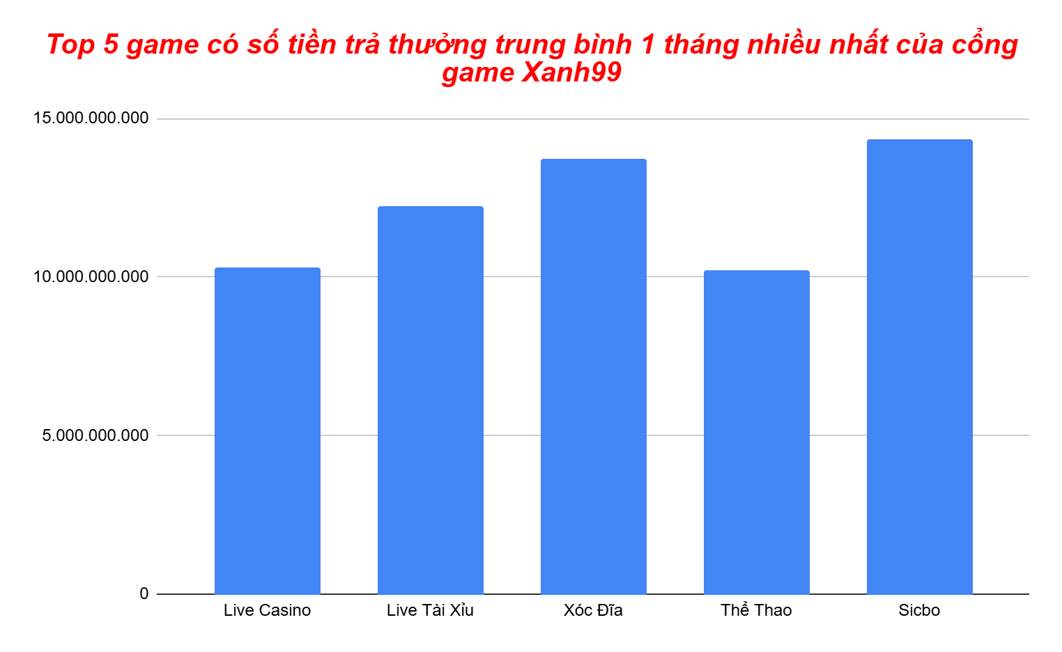
Biểu đồ top 5 game có số tiền chi trả hàng tháng nhiều nhất của Xanh99
Game xóc đĩa xanh99 được trả thưởng trung bình hàng tháng nhiều nhất với số tiền là 13.736.632.138 đ. Game Thể thao được chi trả ít nhất trung bình hàng tháng với số tiền là 10.223.808.312 đ
Zo777 game bài đổi thưởng người chơi đông nhất Việt Nam
Zo777 là một cổng game với lượng người chơi đông đảo nhờ sở hữu kho game đồ sộ, phong phú và hệ thống bảo mật an toàn tuyệt đối, khiến anh em tin tưởng lựa chọn. Khả năng thanh toán linh hoạt và tốc độ xử lý nhanh chóng càng khẳng định độ uy tín của cổng game.
Link tải Zo777
zo777.com/apk
zo777.com/ios
zo777.com/android
zo777.com/webgame
Tên miền chính thức Zo777
zo777.com
zo777.club
zo777.win
zo777.net
Số tiền thắng Zo777 trong năm 2024-2025
Dưới đây là top 5 game mà Zo777 trả thưởng trung bình nhiều nhất theo tháng:
| Game | Số tiền trả thưởng thắng cược trung bình 1 tháng |
| Bài Cào | 10.237.064.013 |
| Tiến Lên Đếm Lá | 14.691.639.989 |
| Bầu Cua | 11.730.907.283 |
| Poker | 12.825.985.810 |
| Tài Xỉu | 9.720.601.614 |
Thống kê top 5 game có số tiền chi trả hàng tháng nhiều nhất của Zo777
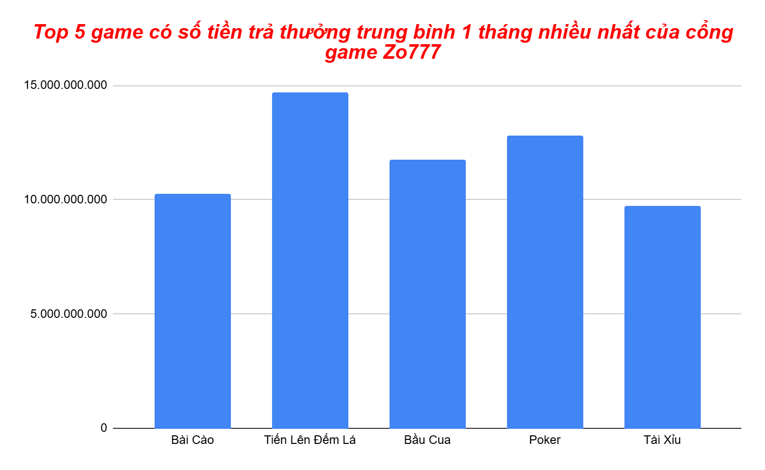
Biểu đồ top 5 game có số tiền chi trả hàng tháng nhiều nhất của Zo777
Game bài tiến lên đếm lá được cổng game Zo777 trả thưởng trung bình hàng tháng nhiều nhất với số tiền 14.691.639.989đ. Tài xỉu là game được chi trả trung bình hàng tháng ít nhất với số tiền 9.720.601.614 đ
1winclub – Cổng game đổi thưởng người chơi đông nhất Việt Nam
1wwinclub được biết đến là cổng game trả thưởng công bằng và minh bạch, với kho game đa dạng và phong phú. Cổng game có hệ thống thanh toán linh hoạt và tốc độ xử lý nhanh chóng, thu hút đông đảo người chơi.
Link tải 1winclub
1winclub.com/apk
1winclub.com/ios
1winclub.com/android
1winclub.com/webgame
Tên miền chính thức 1winclub
1winclub.com
1winclub.club
1winclub.win
1winclub.net
Số tiền thắng 1winclub trong năm 2024-2025
Dưới đây là top 5 game có số tiền trả thưởng trung bình nhiều nhất trong tháng của cổng game 1wwinclub:
| Game | Số tiền trả thưởng thắng cược trung bình 1 tháng |
| Live Xóc Đĩa | 14.401.432.358 |
| Tài Xỉu | 14.565.367.534 |
| Liêng | 11.869.816.290 |
| Mậu Binh | 11.903.493.948 |
| Nổ Hũ | 12.401.425.773 |
Thống kê top 5 game có số tiền chi trả hàng tháng nhiều nhất của 1winclub
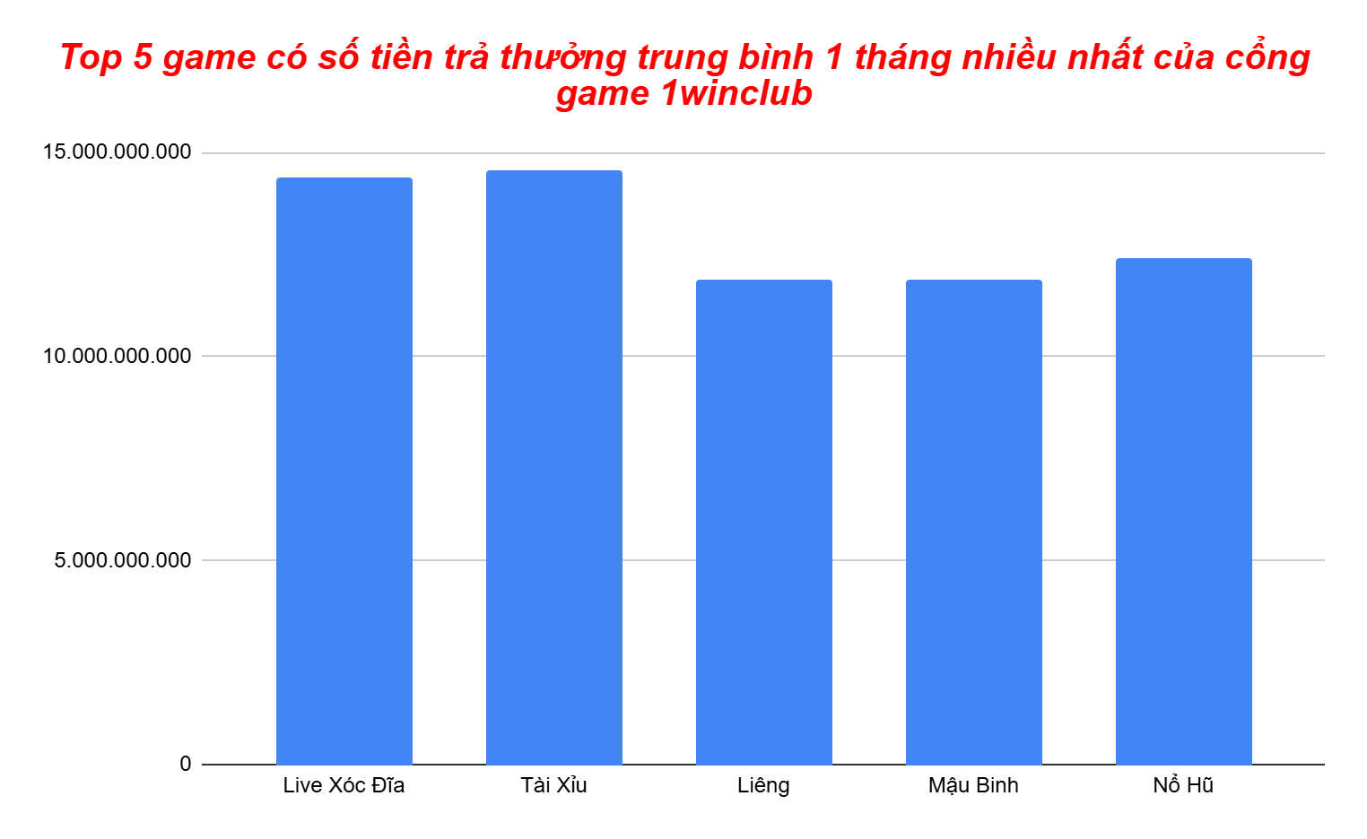
Biểu đồ top 5 game có số tiền chi trả hàng tháng nhiều nhất của 1wwinclub
Tài xỉu có số tiền trả thưởng trung bình hàng tháng là nhiều nhất tại cổng game 1winclub với số tiền là 14.565.367.534 đ. Liêng là game được trả thưởng ít nhất trong bảng thống kê trung bình hàng tháng với số tiền 11.869.816.290 đ
88Kingclub – Cổng game có hội nhóm người chơi đông nhất 2025 – 2024
88Kingclub là một cổng game uy tín với lượng lớn người chơi tham gia. Điểm nổi bật của cổng game là hệ thống chăm sóc khách hàng (CSKH) nhiệt tình và tận tâm. Đặc biệt, 88Kingclub sở hữu hội nhóm Facebook lớn nhất trong làng game đổi thưởng năm 2024. Cổng game còn có hệ thống thanh toán đa dạng và tốc độ xử lý giao dịch nhanh chóng.
Link tải 88kingclub
88kingclub.com/apk
88kingclub.com/ios
88kingclub.com/android
88kingclub.com/webgame
Tên miền chính thức 88kingclub
88kingclub.com
88kingclub.club
88kingclub.win
88kingclub.net
Số tiền thắng 88kingclub trong năm 2024-2025
Dưới đây là top 5 game được anh em chơi nhiều nhất tại 88Kingclub cùng với số tiền trả thưởng mỗi tháng:
| Game | Số tiền trả thưởng thắng cược trung bình 1 tháng |
| Bầu Cua | 11.999.256.217 |
| Mini Poker | 9.917.734.973 |
| Rồng Hổ | 14.279.034.835 |
| Tài Xỉu | 9.140.918.287 |
| Bắn Cá | 9.298.156.779 |
Thống kê top 5 game có số tiền chi trả hàng tháng nhiều nhất của 88Kingclub
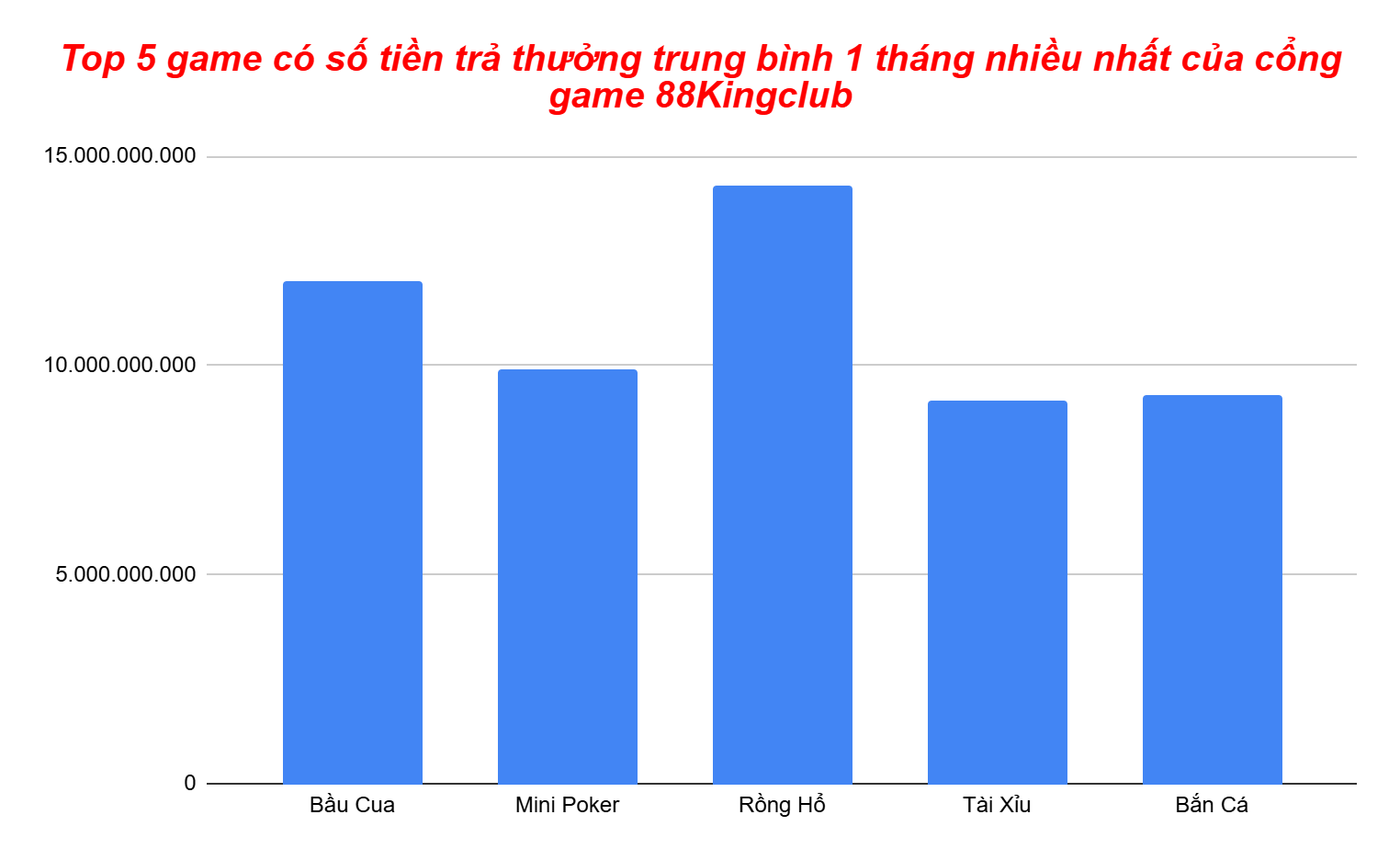
Biểu đồ top 5 game có số tiền chi trả hàng tháng nhiều nhất của 88Kingclub
Rồng hổ có số tiền trả thưởng trung bình hàng tháng là nhiều nhất tại cổng game 88Kingclub với số tiền là 14.279.034.835 đ. Tài Xỉu là game được trả thưởng ít nhất trong bảng thống kê trung bình hàng tháng với số tiền 9.140.918.287 đ
B24club – Cổng game bài đông người chơi nhất 2025 – 2024
B24club là một cổng game bài đổi thưởng mới nhưng đã khẳng định được sự uy tín với giấy phép hoạt động rõ ràng. Cổng game sở hữu kho game phong phú, giao diện mượt mà, và hệ thống thanh toán chất lượng, có khả năng xử lý nhanh chóng các giao dịch lớn.
Link tải B24club
b24club.com/apk
b24club.com/ios
b24club.com/android
b24club.com/webgame
Tên miền chính thức B24club
b24club.com
b24club.club
b24club.win
b24club.net
Số tiền thắng B24club trong năm 2024-2025
Dưới đây là top 5 game có số tiền trả thưởng trung bình hàng tháng nhiều nhất tại B24club:
| Game | Số tiền trả thưởng thắng cược trung bình 1 tháng |
| Nổ Hũ | 10.579.349.439 |
| Lô Đề Siêu Tốc | 11.184.965.747 |
| Thể Thao | 13.670.267.100 |
| Tài Xỉu | 14.979.093.080 |
| Live Sicbo | 14.068.638.680 |
Thống kê top 5 game có số tiền chi trả hàng tháng nhiều nhất của B24club
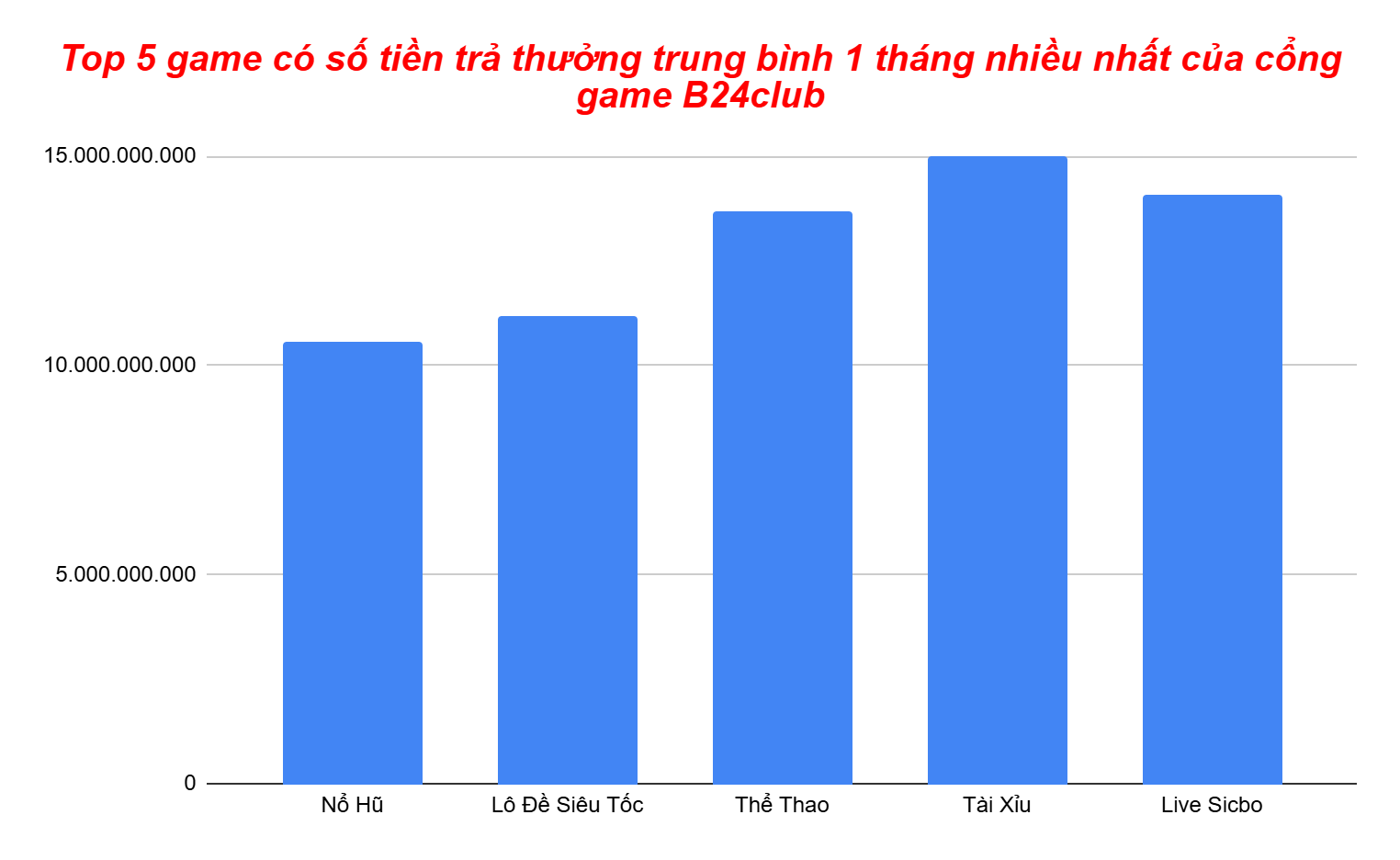
Biểu đồ top 5 game có số tiền chi trả hàng tháng nhiều nhất của B24club
GameTài Xỉu được cổng game B24club trả thưởng trung bình hàng tháng nhiều nhất với số tiền 14.979.093.080đ. Nổ hũ là game được chi trả trung bình hàng tháng ít nhất với số tiền 10.579.349.439 đ
Thông tin bài viết trên lấy từ website: linkgamebaidoithuong.com